
Can a tooth with a large infection be saved? Discover 7 important facts about root canal treatment, microscopic endodontics, and tooth preservation.
Does a Large Tooth Infection Mean You Must Lose Your Tooth?
Imagine visiting your dentist because of persistent tooth pain or swelling. After examining your X-ray, the dentist points to a large dark area surrounding the root of your tooth.
You are told that there is a significant tooth infection and that the tooth might need to be extracted.
Naturally, several questions come to mind.
Can this tooth really be saved? Will root canal treatment work? Is extraction the only option? What happens if the infection spreads?
A tooth infection can be frightening, especially when you see a large dark area on your dental X-ray.
But here’s some encouraging news: a large area of infection or inflammation around a tooth does not automatically mean that the tooth must be removed.
With modern root canal treatment, improved diagnostic imaging, and dental operating microscopes, selected teeth with extensive periapical lesions can be treated successfully.
However, not every tooth can be saved. The outcome depends on the condition of the tooth, the cause of the infection, and several other clinical factors.
If you have been diagnosed with a large tooth infection, understanding these seven important facts can help you make an informed treatment decision.
1. A Large Tooth Infection Around the Root Does Not Always Mean the Tooth Is Beyond Repair
One of the most common misconceptions about dental infections is that the larger the infection, the more likely the tooth needs extraction.
The reality is more complex.
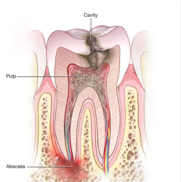
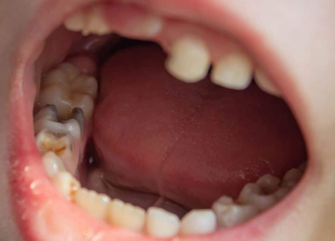
A tooth infection often begins when bacteria enter the dental pulp through deep decay, a crack, trauma, or leakage around an old restoration.
The pulp is the soft tissue inside your tooth that contains nerves, blood vessels, and connective tissue.
When the pulp becomes irreversibly infected or dies, bacteria can multiply within the root canal system.
The body’s immune response to this infection can lead to inflammation and bone loss around the root tip. This condition is known as apical periodontitis.
On a dental X-ray, the affected area may appear as a dark shadow surrounding the root.
What does this dark area mean?
The dark area generally represents a region where normal bone has been lost or altered.
It does not necessarily mean there is a large pocket of pus. Depending on the condition, the lesion may contain inflamed tissue, granulation tissue, or cystic changes.
A large lesion cannot reliably be identified as a cyst or granuloma from its X-ray appearance alone.
The important point is that lesion size is only one part of the diagnosis.
If the tooth has sufficient healthy structure, adequate periodontal support, and no irreparable fracture, root canal treatment may still be a reasonable option.
An examination is necessary before deciding whether extraction is required.
2. Even a Large Tooth Infection May Heal After Proper Root Canal Treatment
Your body has a remarkable ability to repair tissues once the underlying cause of disease is addressed.
A large tooth infection visible on an X-ray does not necessarily require surgical removal of the affected tissue.
In many cases, the infection originates within the root canal system.
If the canals are thoroughly cleaned, disinfected, and sealed, the source of bacterial irritation may be eliminated.
The surrounding tissues can then begin to heal.
How does root canal treatment help?
The treatment involves:
- Accessing the root canal system.
- Removing infected or dead pulp tissue.
- Cleaning and shaping the canals.
- Disinfecting the root canal system.
- Filling and sealing the canals to reduce the risk of reinfection.
- Restoring the tooth to protect it against leakage and fracture.
Over time, the bone surrounding the root may gradually regenerate.
Large lesions can sometimes show substantial healing following nonsurgical endodontic treatment, although healing is not guaranteed.
Published case reports have documented the healing of extensive periapical lesions following root canal treatment or retreatment.
3. The Condition of Your Tooth Matters More Than the Size of the Infection Alone
When evaluating whether a tooth can be saved, the dentist must consider more than the X-ray appearance.
To determine whether a tooth infection can be managed successfully, several important questions must be answered.
Is there enough healthy tooth structure remaining?
A severely decayed or broken tooth may not have sufficient structure to support a long-term restoration.
Even if the infection is eliminated, the tooth must be strong enough to function during chewing.
Is there a vertical root fracture?
A vertical root fracture can make a tooth extremely difficult or impossible to preserve predictably.
What is the condition of the surrounding gums and bone?
Advanced periodontal disease and severe loss of tooth support can affect the long-term prognosis.
Can the root canals be adequately treated?
The anatomy of the root canals, previous treatment, existing posts, and procedural complications can influence treatment planning.
Can the tooth be properly restored?
A reliable final restoration is essential to reduce bacterial leakage and protect the remaining tooth structure.
The objective is not simply to eliminate the infection. It is to preserve a tooth that can remain comfortable and functional over the long term.
4. A Dental Operating Microscope Can Help Treat Complex Root Canal Problems
Modern endodontic treatment has advanced significantly with the use of magnification, improved instruments, and enhanced imaging.
When a tooth infection begins inside a complex root canal system, effective treatment may require identifying and managing anatomical details that are difficult to see with the naked eye.
Some teeth have additional canals, narrow passages, unusual root anatomy, or obstructions from previous treatment.
These factors may complicate the treatment.
How does a dental operating microscope help?
A dental operating microscope provides magnification and illumination during treatment.
This can help the dentist:
- Identify additional or previously missed canals.
- Locate narrow or calcified canal openings.
- Examine the internal tooth structure more closely.
- Manage selected complications from previous treatment.
- Perform precise procedures while aiming to preserve healthy tooth structure.
Microscopes are particularly useful in complex retreatment cases, where previous filling materials must be removed and the canals reassessed.
At Joglekar’s Dental Solutions, Chinchwad, we use dental operating microscopes for precision-focused endodontic treatments.
However, magnification alone does not guarantee treatment success. Appropriate diagnosis, disinfection, sealing, and final restoration remain essential.
5. What If a Previously Root-Canal-Treated Tooth Develops a Large Infection?
Have you ever undergone root canal treatment only to discover an infection around the same tooth months or years later?
This can be disappointing, particularly if you were told that the tooth had already been treated.
But a previously treated tooth does not always need extraction.
If a tooth infection remains or develops after root canal treatment, the dentist must determine the underlying cause.
Possible reasons include missed canals, persistent microorganisms, inadequate sealing, new decay, or leakage around an old crown.
Occasionally, a fracture or another problem may be responsible.
Option 1: Root canal retreatment
During retreatment, the dentist reopens the tooth, removes the previous root canal filling, and examines the canal system.
The canals are then cleaned, disinfected, and sealed again.
A new restoration may be needed afterward.
Option 2: Endodontic microsurgery
If nonsurgical retreatment is impractical or the lesion persists, endodontic surgery may be considered for suitable cases.
A procedure called an apicoectomy involves accessing the root tip surgically, removing the affected root-end tissue and a small portion of the root, and sealing the root end when indicated.
Option 3: Extraction and replacement
If the tooth is structurally unrestorable, has an irreparable fracture, or cannot be maintained predictably, extraction may be necessary.
Replacement options can then be discussed.
Retreatment, surgery, and extraction are different options, not interchangeable solutions for every patient. The appropriate choice depends on the diagnosis and expected prognosis.